Verticillium Wilt: Difference between revisions
No edit summary |
|||
| Line 12: | Line 12: | ||
When a cross section is taken, infected branches on trees will show dark green or brown rings[1]. This is because verticillium infects and spreads through the vascular systems of its hosts, this disrupts the water and mineral transportation to the branches and leaves of the hosts[3]. While vascular staining occurs often, it is not always present[3]. | When a cross section is taken, infected branches on trees will show dark green or brown rings[1]. This is because verticillium infects and spreads through the vascular systems of its hosts, this disrupts the water and mineral transportation to the branches and leaves of the hosts[3]. While vascular staining occurs often, it is not always present[3]. | ||
=Life Cycle= | =Life Cycle= | ||
''V. dahliae'' | ''V. dahliae'' exists in three life stages; dormant, parasitic, and saprophytic. | ||
====Dormant==== | ====Dormant==== | ||
In its dormant phase, mycelia and microsclerotia (dark, durable, resting structures produced by the fungus[8])of the fungus are capable of surviving in dried conditions, they can survive in soil away from a host or embedded in fragments of tissue[6]. The structures will be ready to germinate once in the presence of a host[7]. The microsclerotia can either stand alone, become embedded in plant leaves or branches; once the branches or leaves die and fall off the tree they can be transported by wind to other places. Because | In its dormant phase, mycelia and microsclerotia (dark, durable, resting structures produced by the fungus[8])of the fungus are capable of surviving in dried conditions, they can survive in soil away from a host or embedded in fragments of tissue[6]. The structures will be ready to germinate once in the presence of a host[7]. The microsclerotia can either stand alone, become embedded in plant leaves or branches; once the branches or leaves die and fall off the tree they can be transported by wind to other places[3]. Because microsclerotia allow verticillium to lie dormant for a long time, it is highly unlikely that the soil it is infecting will ever be rid of the fungus. The only way to avoid infecting plant are to plant species that are not susceptible to infection by the fungus[3]. | ||
====Parasitic==== | ====Parasitic==== | ||
Revision as of 17:54, 2 May 2019

Verticillium wilt is the result of a soil-borne fungal pathogen called Verticillium dahliae that infects over 200 species of plants[1]. In all there are ten recognized species of Verticillium but V. dahliae has the widest range of hosts. These can include, maple trees, tomato plants, eggplants, peach trees, black raspberry, spinach, pumpkin, alfalfa, hops, cherry trees, peony, snapdragons, chrysanthemums, etc[1]. As said there are over 200 species that acts as a host, many of which include many important agriculture crops, or forest species.
Signs and Symptoms
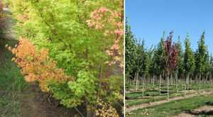
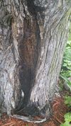
There are many signs and symptoms of Verticillium Wilt that a host plant may exhibit. These include, wilting of the leaves, chlorosis (yellowing of the leaves), stunted plant growth[1], the edges of the leaves my appear "scorched" or brown, and dead twigs and branches may appear. Specifically on maples areas of dead bark, called cankers, may appear[2].

These symptoms may appear on one side of the plant as a whole, one branch, or one grouping of leaves. Symptoms are most nocticble from mid to late summer or during times of extreme heat or drought[1].


Symptoms expressed are dependent on the host for example, in spinach or cauliflower symptoms don't appear until the plant begins to flower[1].
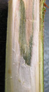
When a cross section is taken, infected branches on trees will show dark green or brown rings[1]. This is because verticillium infects and spreads through the vascular systems of its hosts, this disrupts the water and mineral transportation to the branches and leaves of the hosts[3]. While vascular staining occurs often, it is not always present[3].
Life Cycle
V. dahliae exists in three life stages; dormant, parasitic, and saprophytic.
Dormant
In its dormant phase, mycelia and microsclerotia (dark, durable, resting structures produced by the fungus[8])of the fungus are capable of surviving in dried conditions, they can survive in soil away from a host or embedded in fragments of tissue[6]. The structures will be ready to germinate once in the presence of a host[7]. The microsclerotia can either stand alone, become embedded in plant leaves or branches; once the branches or leaves die and fall off the tree they can be transported by wind to other places[3]. Because microsclerotia allow verticillium to lie dormant for a long time, it is highly unlikely that the soil it is infecting will ever be rid of the fungus. The only way to avoid infecting plant are to plant species that are not susceptible to infection by the fungus[3].
Parasitic
Interactions with Arbuscular Mycorrhizal Fungi
References
[1]Dung, Jeremiah K.S., and Jerry Weiland. “Verticillium Wilt in the Pacific Northwest.” Pacific Northwest Pest Management Handbooks, OSU Extension Service - Extension and Experiment Station Communications, 13 Oct. 2016, pnwhandbooks.org/plantdisease/pathogen- articles/common/fungi/verticillium-wilt-pacific-northwest.
[2]“Verticillium Wilt.” Verticillium Wilt | The Morton Arboretum, www.mortonarb.org/trees-plants/tree-and-plant-advice/help- diseases/verticillium-wilt.
[3]Brazee, Nicholas. “Verticillium Wilt.” Center for Agriculture, Food and the Environment, 26 Feb. 2018, ag.umass.edu/landscape/fact- sheets/verticillium-wilt.
[4]Anita. “Silver Maple - Bleeding Canker? - Ask an Expert.” EXtension, 14 June 2017, ask.extension.org/questions/406833.
[5]Gubler, W D, and B L Teviotdale. “How to Manage Pests.” UC IPM Online, University of California, ipm.ucanr.edu/PMG/r602101511.html.
[6]Schnathrost, W C. “Fungal Wilt Diseases of Plants.” Fungal Wilt Diseases of Plants, by Carl H. Beckman et al., Academic., 2012, pp. 81–108.
[7]Inderbitzin, Patrik, et al. “Phylogenetics and Taxonomy of the Fungal Vascular Wilt Pathogen Verticillium, with the Descriptions of Five New Species.” PloS One, Public Library of Science, 7 Dec. 2011, www.ncbi.nlm.nih.gov/pmc/articles/PMC3233568/.
[8]Gordee, R. S., and C. L. Porter. “Structure, Germination, and Physiology of Microsclerotia of Verticillium Albo-Atrum.” Mycologia, vol. 53, no. 2, 1961, pp. 171–182. JSTOR, www.jstor.org/stable/3756235.